Your visit · 5 min read
Panoramic X-ray or 3D Scan? What Each One Shows and When You Need It
A panoramic X-ray shows your whole mouth in one flat image. A 3D cone-beam scan shows depth. What each one is for, how they differ in radiation, and when a dentist needs one or the other.
Mineers Smile CenterReviewed by Dr. Elias Abou Tayeh, DDS
Your dentist says you need a panoramic X-ray, or a 3D scan, and you wonder whether they are the same thing and why one might be chosen over the other. They are not the same. One is a single wide picture of your whole mouth. The other is a three-dimensional model. Here is what each one shows, how they compare, and when a dentist needs which.
The panoramic X-ray
A panoramic X-ray shows your entire mouth in one image. Cleveland Clinic says it shows teeth, jaws, nerves, sinuses and bone (Cleveland Clinic). It is a flat, two-dimensional picture, taken while the machine moves around your head.
Dentists use it to get an overview: how teeth are positioned, where wisdom teeth sit, the state of the jaw bones, and signs of infection that cannot be seen by looking. It is not the usual tool for fine detail on a single tooth. For that, dentists use close-up images such as bitewing X-rays, which Cleveland Clinic says show cavities between teeth and under the gum line, and periapical X-rays, which detect gum disease, bone loss and cavities near tooth roots.
The 3D scan (cone-beam CT)
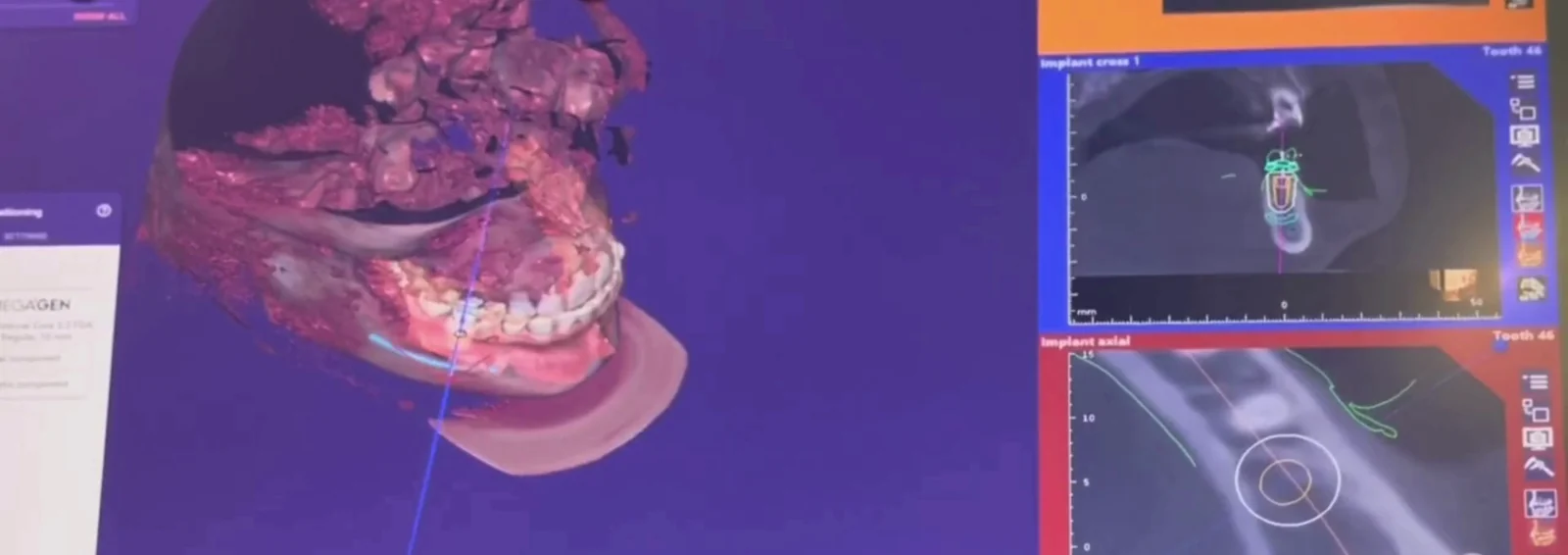
A cone-beam computed tomography (CBCT) scan uses a cone-shaped X-ray beam that rotates around you. The US Food and Drug Administration (FDA) says the data are used to reconstruct a three-dimensional image of the teeth, the mouth and jaw region, and nearby structures, where a conventional X-ray gives two-dimensional information (FDA).
The FDA lists what dentists use CBCT for: dental implant planning, looking at abnormal teeth, evaluating the jaws and face, diagnosing cavities, root canal diagnosis and dental trauma. Cleveland Clinic says it is often used for implant planning because it shows teeth, jaws, nerves and sinuses in three dimensions.
How they compare
| Panoramic X-ray | 3D cone-beam scan | |
|---|---|---|
| Image | One flat picture of the whole mouth | A three-dimensional model |
| Shows | Teeth, jaws, nerves, sinuses, bone | The same structures with depth |
| Often used for | An overview, wisdom teeth, general checks | Implant planning, root canal diagnosis, complex cases |
| Radiation | A small dose | The FDA says CBCT typically gives more than a conventional dental X-ray, though generally less than other CT exams |
Neither is "better". A panoramic image answers a general question quickly. A 3D scan answers a more specific one that a flat image cannot, such as how deep the bone is where an implant will go.
When do you need one?
The American Dental Association and the FDA say dental X-rays, including CBCT, should be taken only when needed for the diagnosis or treatment of disease. The FDA's advice for patients is simple: ask whether the exam is necessary, whether there are equally useful alternatives that use less or no radiation, and keep a record of your imaging history so exams are not repeated for no reason.
For routine care, Cleveland Clinic says people with healthy teeth and gums usually need new X-rays every year, and may need them more often if they are prone to cavities or gum disease. For specific plans:
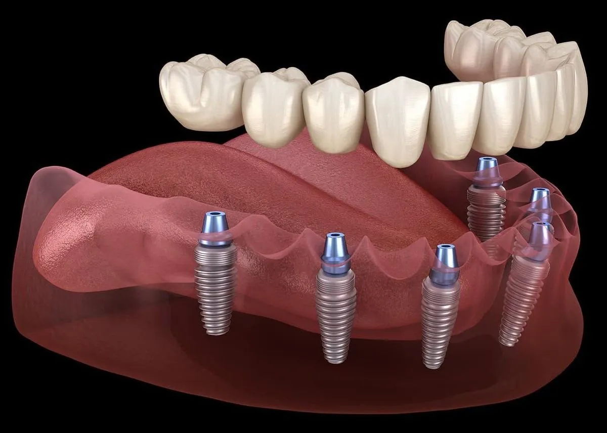
- Implants. A 3D scan shows how much bone you have and where the nerves run, so the implant can be planned to sit safely. Single Implant, Several Teeth or a Full Arch? explains how that plan is made.
- Braces and aligners. Dentists may use X-rays to plan treatment. Aligners also need a scan of the teeth, but that is different, see below.
- A tooth that hurts. An X-ray helps show an infection at the root. Toothache: What to Do Tonight covers when to call.
A 3D scan of your teeth is not always an X-ray
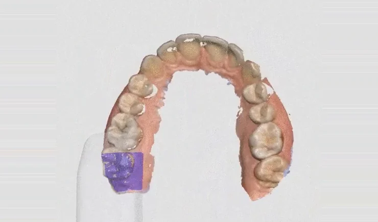
At Mineers, "3D" can mean two things. A 3D X-ray of your jaw is used to plan implants and shows bone and nerves. A 3D digital scan of your teeth is made with a small handheld scanner that records the surface of your teeth, and it is what clear aligners and many restorations are planned from. It is an optical scan, not an X-ray. The aligner page says it takes one visit, with no trays of putty in your mouth.
Safety questions
- Radiation. Cleveland Clinic says dental X-rays use a tiny amount of radiation, and that digital X-rays use 80% to 90% less radiation than traditional film. A lead apron and thyroid collar may be used.
- Pregnancy. Cleveland Clinic says dental X-rays are generally considered safe in pregnancy and breastfeeding, that the risk to the fetus is minimal, and that most dentists avoid them unless absolutely necessary.
- Children. The FDA says concerns about radiation are greater for younger patients because they are more sensitive to it.
Bringing an X-ray with you
If you already have a recent X-ray, bring the image or the file. It helps the team plan and can avoid repeating an exam. If you live abroad, How to Send a Photo of Your Smile explains what to send. A recent X-ray sent by WhatsApp lets us tell you which treatment may suit you, and your plan is confirmed after an in-person examination.
Frequently Asked Questions
What is the difference between a panoramic X-ray and a CBCT scan?
A panoramic X-ray is a single flat image of your whole mouth. A CBCT scan creates a three-dimensional image, which is why it is often used to plan implants.
Is a panoramic X-ray enough to plan an implant?
It gives a useful overview, but implants are usually planned on a 3D scan, because it shows the bone and nerves in three dimensions. At Mineers the implant is planned on a 3D X-ray.
Is a dental X-ray safe?
Cleveland Clinic says dental X-rays use a tiny amount of radiation, and the FDA says the clinical benefit of an appropriate exam outweighs the small risk. They should only be taken when needed.
How often do I need a panoramic X-ray?
It depends on your teeth and your plan. Ask your dentist why one is needed and how long the last one stays useful, and keep copies of your images.
Can I use an X-ray from another dentist?
Often, if it is recent and clear. Bring or send the file, and the team will tell you if a new one is needed.
Planning treatment and not sure what you need? Book a private consultation, or send us a note on WhatsApp, +961 3 757 127.
This article is general information, not a diagnosis. The right images for you are decided by your dentist.
Independent sources
Where to read more, from independent health organisations.